
Electromiografía
Laboratorio de Fisiología Oral, DEPeI Odontología, UNAM
Dr. Luis Antonio García Espinosa
| Ciudad de México | septiembre 19, 2022 |
Contenido
- Generación de señal EMGS
- Extracción de señal EMGS
- Extracción de información útil
- Buenas prácticas de registro de EMGS
Generación de señal EMGS
Generación de señal EMGS
Generación de señal EMGS
Extracción de la señal EMGS
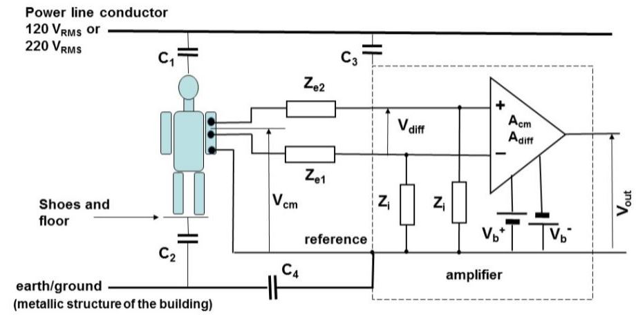
Representación esquemática de un amplificador de EMGS canal simple conectado a un sujeto (Merletti, 2020)
Extracción de la señal EMGS
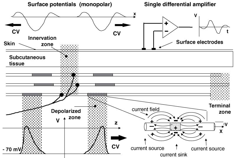
Representación de una unidad motora (UM) y un potencial de acción de unidad motora [26]
El análisis del proceso de generación y propagación del AP lleva a las siguientes conclusiones importantes:
- Las características de la señal EMGS son influenciadas significativamente por factores distintos a los de mayor interés para el estudio de procesos fisiológicos del sistema neuromuscular.
- Cambios en el conductor de volumen, la colocación de los electrodos de superficie, pueden generar cambios en las características de la señal EMGS similares a los generados por procesos fisiológicos de interés. Esta situación, dificulta su interpretación y asociación con procesos fisiológicos de mayor interés.
- Es indispensable definir las mejores prácticas en el uso de la EMGS para fines clínicos para mitigar los factores de variabilidad indeseados que pueden llevar a interpretaciones erróneas sobre el sistema neuromuscular.
- Es necesario explorar nuevos enfoques de análisis de señal EMGS que sean menos sensibles a fuentes de variabilidad indeseadas.
Extracción de la señal EMGS
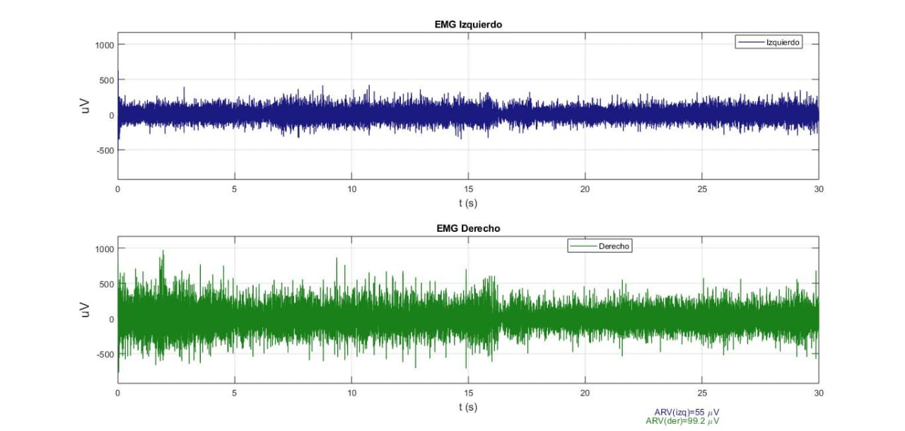
Señal EMGS cruda registrada durante contracción isométrica en oclusión centrica (OC), (Laboratorio de fisiología oral, 2019)
Extracción de información útil
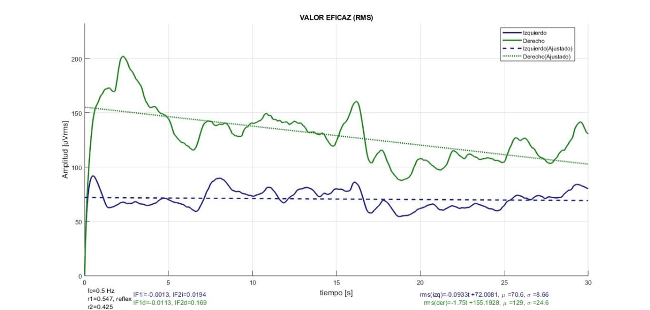
Valor RMS de señal EMGS cruda registrada durante contracción isométrica en oclusión centrica (OC), (Laboratorio de fisiología oral, 2019)
Extracción de información útil
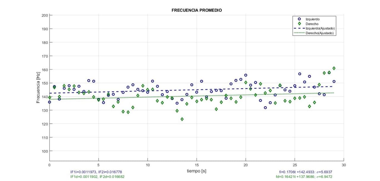
Valor de la frecuencia promedio MNF (Mean Frequency) de señal EMGS cruda registrada durante contracción isométrica en oclusión centrica (OC), (Laboratorio de fisiología oral, 2019)
Extracción de información útil

Indice de Hurts de señal EMGS cruda registrada durante contracción isométrica en oclusión centrica (OC), (Laboratorio de fisiología oral, 2019)
Mejores prácticas
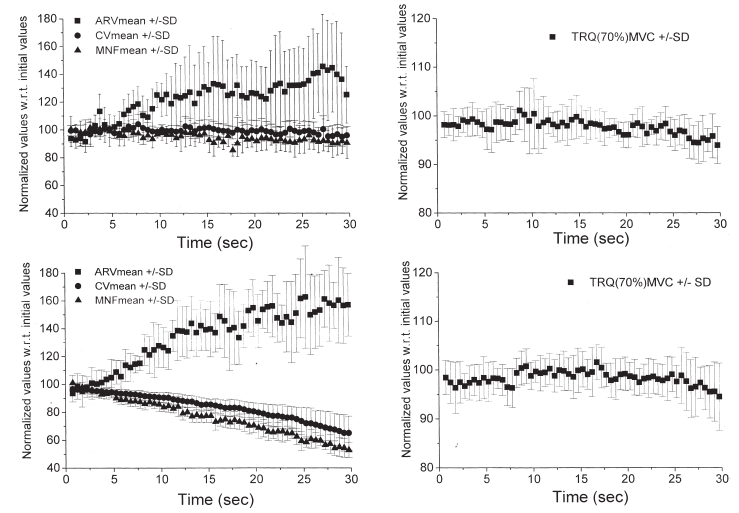
Ejemplos de gráficas de fatiga al 70% de máxima contracción voluntaria (MCV) de músculo bíceps braquial de dos sujetos [51]
Las aplicaciones clínicas de EMGS demandan confiabilidad y repetibilidad de las técnicas utilizadas para el diagnóstico y seguimiento de los pacientes [10].
La repetibilidad de las variables extraídas de la señal EMGS basadas en la estimación de la amplitud y la densidad de la potencia espectral, como RMS, ARV, MNF y MDF, mejora si se utiliza una metodología de estandarización y normalización como la sugerida por [26-30]. Sin embargo, no siempre es viable.
Mejores prácticas
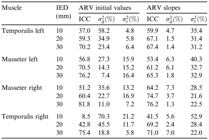
Coeficiente de correlación intraclase (ICC) para el valor inicial y pendiente de la variable ARV extraída de señal EMGS de cuatro músculos de las masticación [26]
La repetibilidad de la variable ARV dentro de cada sujeto y entre sujetos mejora al aumentar la distancia inter-electrodos (IED). Pero es aplicable solo si se realiza contracción isométrica controlada a través de un sistema de bioretroalimentación que incluya un sensor de fuerza de mordida.
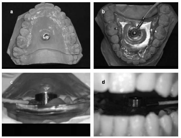
Sensor de fuerza usado por Castroforio et al. para evaluar la reproducibilidad de las variables EMGS de los músculo elevadores de la mandíbula [26].
Mejores prácticas
- Entrenamiento en la colocación de electrodos superficiales.
- No registrar EMGS en contracción que genere fatiga conciente en el paciente.
Referencias
[1] G. M. Tartaglia, G. Lodetti, G. Paiva, C. M. D. Felicio, y C. Sforza, “Surface elec-tromyographic assessment of patients with long lasting temporomandibular joint disor-der pain”, Journal of Electromyography and Kinesiology, vol. 21, pp. 659–664, 2011, doi: https://doi.org/10.1016/j.jelekin.2011.03.003.
[2] M. R. R. Fernando Ángeles Medina, Dolor Orofacial y Desórdenes de la Articulación Temporomandibular, 1a ed. Trillas, 2006.
[3] J. M. Axel Bumann Ulrich Lotzmann, TMJ Disorders and Orofacial Pain The Role of Den-tistry in a Multidisciplinary Diagnostic Approach. Stuttgart, Germany: Thieme, 2002.
[4] G. O. Kruger, Cirugía buco-maxilofacial, 5a ed. Panamericana, 1983.
[5] V. Kasat, A. Gupta, R. Ladda, M. Kathariya, H. Saluja, y A.-A. Farooqui, “Transcutaneous electric nerve stimulation (TENS) in dentistry- A review”, Journal of clinical and experi-mental dentistry, vol. 6, núm. 5, pp. e562–e568, dic. 2014, doi: 10.4317/jced.51586.
[6] Y.-C. H. Chun-En Aurea Kuo Szu-Ying Wu y W.-L. Hu, “The Application and Effectiveness of Electroacupuncture in the Pain Management of Temporomandibular Disorders”, en Acupuncture, SM Group, 2017. [En línea]. Disponible en: https://smjournals.com/ebooks/acupuncture/index.php
[7] R.-B. D. Gomes NC Berni-Schwarzenbeck KC, Packer AC, “Effect of cathodal high-voltage electrical stimulation on pain in women with TMD”, Rev Bras Fisioter, vol. 16, núm. 1, pp. 10–5, feb. 2012.
[8] G. D. Klasser y J. P. Okeson, “The clinical usefulness of surface electromyography in the diagnosis and treatment of temporomandibular disorders”, The Journal of the American Dental Association, vol. 137, pp. 763–771, 2006, doi: https://doi.org/10.14219/jada.archive.2006.0288.
[9] U. Santana-Mora et al., “Changes in EMG activity during clenching in chronic pain pa-tients with unilateral temporomandibular disorders”, Journal of Electromyography and Kinesiology, vol. 19, pp. e543–e549, 2009, doi: https://doi.org/10.1016/j.jelekin.2008.10.002.
Referencias
[10] D. F. Roberto Merletti, Surface Electromyography, Physiology, Engineering, and Applica-tions. United States of America: IEEE Press, 2016.
[11] B. C. Moffett Jr., L. C. Johnson, J. B. McCabe, y H. C. Askew, “Articular remodeling in the adult human temporomandibular joint”, American Journal of Anatomy, vol. 115, núm. 1, pp. 119–141, 1964, doi: https://doi.org/10.1002/aja.1001150108.
[12] S. A. Lévano Loayza y A. T. Sovero Gaspar, “Evaluación anatómica de la articulación temporomandibular mediante resonancia magnética. Artículo de revisión”, Revista Es-tomatológica Herediana, vol. 30, núm. 4, pp. 285–293, ene. 2021, doi: 10.20453/reh.v30i4.3882.
[13] T. Castroflorio, D. Falla, G. M. Tartaglia, C. Sforza, y A. Deregibus, “Myoelectric mani-festations of jaw elevator muscle fatigue and recovery in healthy and TMD subjects”, Journal of Oral Rehabilitation, vol. 39, pp. 648–658, 2012, doi: 10.1111/j.1365-2842.2012.02309.x.
[14] P. Rosenfalck, “Intra- and extracellular potential fields of active nerve and muscle fi-bres. A physico-mathematical analysis of different models”, Acta physiologica Scandina-vica. Supplementum, vol. 321, p. 1—168, 1969.
[15] R. Merletti, L. Lo Conte, E. Avignone, y P. Guglielminotti, “Modeling of surface myoelec-tric signals. I. Model implementation”, IEEE Transactions on Biomedical Engineering, vol. 46, núm. 7, pp. 810–820, 1999, doi: 10.1109/10.771190.
[16] L. A. García-Espinosa et al., “Multi-fractal DFA analysis of masseter muscles SEMG sig-nal in patients with TMD, pilot study”, Biomedical Signal Processing and Control, vol. 68, p. 102732, 2021, doi: https://doi.org/10.1016/j.bspc.2021.102732.
[17] C.-K. Peng, S. V. Buldyrev, S. Havlin, M. Simons, H. E. Stanley, y A. L. Goldberger, “Mosa-ic organization of DNA nucleotides”, Phys. Rev. E, vol. 49, núm. 2, pp. 1685–1689, feb. 1994, doi: 10.1103/PhysRevE.49.1685.
Referencias
[18] C. K. Peng, S. Havlin, H. E. Stanley, y A. L. Goldberger, “Quantification of scaling expo-nents and crossover phenomena in nonstationary heartbeat time series”, Chaos: An In-terdisciplinary Journal of Nonlinear Science, vol. 5, pp. 82–87, 1995, doi: 10.1063/1.166141.
[19] J. Walleczek, Self-Organized Biological Dynamics and Nonlinear Control. New York: Cambridge University Press, 2000.
[20] H. E. Hurst, “Long-Term Storage Capacity of Reservoirs”, Transactions of the American Society of Civil Engineers, vol. 116, núm. 1, pp. 770–799, 1951, doi: 10.1061/TACEAT.0006518.
[21] A. N. Kolmogorov, “The Local Structure of Turbulence in Incompressible Viscous Fluid for Very Large Reynolds Numbers”, Proceedings: Mathematical and Physical Sciences, vol. 434, núm. 1890, pp. 9–13, 1991.
[22] J. Feder, Fractals, 1a ed. Boston, MA: Springer, 1988. doi: 10.1007/978-1-4899-2124-6.
[23] J. W. Kantelhardt, S. A. Zschiegner, E. Koscielny-Bunde, S. Havlin, A. Bunde, y H. E. Stan-ley, “Multifractal detrended fluctuation analysis of nonstationary time series”, Physica A Statistical Mechanics and its Applications, vol. 316, núm. 1, pp. 87–114, dic. 2002, doi: 10.1016/S0378-4371(02)01383-3.
[24] T. Castroflorio, D. Farina, A. Bottin, M. G. Piancino, P. Bracco, y R. Merletti, “Surface EMG of jaw elevator muscles: Effect of electrode location and inter-electrode distance”, Journal of Oral Rehabilitation, vol. 32, pp. 411–417, 2005, doi: 10.1111/j.1365-2842.2005.01442.x.
[25] P. Ravier, O. Buttelli, R. Jennane, y P. Couratier, “An EMG fractal indicator having dif-ferent sensitivities to changes in force and muscle fatigue during voluntary static mus-cle contractions”, Journal of Electromyography and Kinesiology, vol. 15, pp. 210–221, 2005, doi: https://doi.org/10.1016/j.jelekin.2004.08.008.
Referencias
[26] T. Castroflorio et al., “Reproducibility of surface EMG variables in isometric sub-maximal contractions of jaw elevator muscles”, Journal of Electromyography and Kine-siology, vol. 16, pp. 498–505, 2006, doi: https://doi.org/10.1016/j.jelekin.2005.08.007.
[27] C. M. D. E. Felício, F. V. Sidequersky, G. M. Tartaglia, y C. Sforza, “Electromyographic standardized indices in healthy Brazilian young adults and data reproducibility”, Journal of Oral Rehabilitation, vol. 36, pp. 577–583, 2009, doi: 10.1111/j.1365-2842.2009.01970.x.
[28] V. F. Ferrario, C. Sforza, A. Colombo, y V. Ciusa, “An electromyographic investigation of masticatory muscles symmetry in normo-occlusion subjects”, Journal of Oral Rehabilita-tion, vol. 27, pp. 33–40, 2000, doi: 10.1046/j.1365-2842.2000.00490.x.
[29] V. F. Ferrario, G. M. Tartaglia, A. Galletta, G. P. Grassi, y C. Sforza, “The influence of occlusion on jaw and neck muscle activity: a surface EMG study in healthy young adults”, Journal of Oral Rehabilitation, vol. 33, pp. 341–348, 2006, doi: https://doi.org/10.1111/j.1365-2842.2005.01558.x.
[30] S. E. Forrester, S. J. Allen, R. G. Presswood, C. T. O. Y. A., y G. P. A. I. N. M. T., “Neuro-muscular function in healthy occlusion”, Journal of Oral Rehabilitation, vol. 37, pp. 663–669, 2010, doi: https://doi.org/10.1111/j.1365-2842.2010.02097.x.
[31] D. K. Kumar, S. P. Arjunan, y B. Aliahmad, FRACTALS: Applications in Biological Signal-ling and Image Processing. Boca, Raton, FL: CRC Press, 2017.
[32] A. G. P. J. S. Bendat, Random data: Analysis and Measurement Procedures. New Jersey: Wiley, 2010.
[33] C. J. Anmuth, G. Goldberg, y N. H. Mayer, “Fractal dimension of electromyographic sig-nals recorded with surface electrodes during isometric contractions is linearly correlat-ed with muscle activation”, Muscle & Nerve, vol. 17, pp. 953–954, 1994, doi: 10.1002/mus.880170819.
[34] T. Nguyen-Ky, P. Wen, y Y. Li, “Improving the accuracy of depth of anaesthesia using modified detrended fluctuation analysis method”, Biomedical Signal Processing and Control, vol. 5, pp. 59–65, 2010, doi: https://doi.org/10.1016/j.bspc.2009.03.001.
Referencias
[35] J. B. Gao, C. Yinhe, W. Ten, y H. U. J., Multiscale analysis of complex time series. New Jersey: Wiley Press, 2007.
[36] M. Talebinejad, A. D. C. Chan, A. Miri, y R. M. Dansereau, “Fractal analysis of surface electromyography signals: A novel power spectrum-based method”, Journal of Electro-myography and Kinesiology, vol. 19, pp. 840–850, 2009, doi: https://doi.org/10.1016/j.jelekin.2008.05.004.
[37] M. Talebinejad, A. D. C. Chan, y A. Miri, “Fatigue estimation using a novel multi-fractal detrended fluctuation analysis-based approach”, Journal of Electromyography and Kine-siology, vol. 20, pp. 433–439, 2010, doi: https://doi.org/10.1016/j.jelekin.2009.06.002.
[38] R. Merletti y G. L. Cerone, “Tutorial. Surface EMG detection, conditioning and pre-processing: Best practices”, Journal of Electromyography and Kinesiology, vol. 54, p. 102440, 2020, doi: https://doi.org/10.1016/j.jelekin.2020.102440.
[39] L. A. García-Espinosa, “Diseño y construcción de electromiógrafo para el registro de EMG superficial de músculos maseteros e implementación del análisis multifractal por DFA”, Centro de Investigación y de Estudios Avanzados del Intituto Politécnico Nacional (CINVESTAV), México, D.F., 2011.
[40] L. de Fisiología F.O. UNAM, “Manual de usuario electromiógrafo digital y medidor de fuerza de mordida. PAPIIT IN227511”.
[41] “Standards for Reporting EMG Data”, Journal of Electromyography and Kinesiology, vol. 42, p. I–II, 2018, doi: https://doi.org/10.1016/S1050-6411(18)30348-1.
[42] C. I. Rodríguez-Castañeda et al., “Cambios de la actividad electromiográfica durante las diferentes fases del tratamiento de ortodoncia: resultados de una prueba piloto”, Revis-ta Mexicana de Ortodoncia, vol. 5, pp. 238–244, 2017, doi: https://doi.org/10.1016/j.rmo.2018.01.006.
[43] C. I. Rodríguez-Castañeda et al., “Changes of EMG Activity During Orthodontic Treat-ment: Cohort Study”, San Francisco California, Francisco, California, 2017.
Referencias
[44] P. A. P. R. Merletti, Electromyography Physiology, Engineering, and Noninvasive Appli-cations. United States of America: Wiley-Interscience, 2004.
[45] M. Naeije, R. S. McCarroll, y W. A. Weijs, “Electromyographic activity of the human masticatory muscles during submaximal clenching in the inter-cuspal position.”, Journal of Oral Rehabilitation, vol. 16, p. 63, 1989.
[46] E. A. Clancy, E. L. Morin, y R. Merletti, “Sampling, noise-reduction and amplitude esti-mation issues in surface electromyography”, Journal of Electromyography and Kinesiol-ogy, vol. 12, pp. 1–16, 2002, doi: https://doi.org/10.1016/S1050-6411(01)00033-5.
[47] R. Merletti, M. Knaflitz, y C. J. D. Luca, “Myoelectric manifestations of fatigue in volun-tary and electrically elicited contractions”, Journal of Applied Physiology, vol. 69, pp. 1810–1820, 1990, doi: 10.1152/jappl.1990.69.5.1810.
[48] M. H. Hayes, Statistical Digital Signal Processing and Modeling. United States of Ameri-ca: John Wiley & Sons, Inc, 1996.
[49] J. G. W. Ramón Pallás-Areny, Analog Signal Processing, 1a ed. Wiley, 1999.
[50] J. Gomez-Clapers, E. Serrano-Finetti, R. Casanella, y R. Pallas-Areny, “Can driven-right-leg circuits increase interference in ECG amplifiers?”, en 2011 Annual International Conference of the IEEE Engineering in Medicine and Biology Society, 2011, pp. 4780–4783. doi: 10.1109/IEMBS.2011.6091184.
[51] A. Rainoldi, G. Galardi, L. Maderna, G. Comi, L. L. Conte, y R. Merletti, “Repeatability of surface EMG variables during voluntary isometric contractions of the biceps brachii muscle”, Journal of Electromyography and Kinesiology, vol. 9, pp. 105–119, 1999, doi: https://doi.org/10.1016/S1050-6411(98)00042-X.
[52] L. S. Moreira, L. A. Elias, C. M. Germer, y E. T. Palomari, “Reliable measurement of in-cisal bite force for understanding the control of masticatory muscles”, Archives of Oral Biology, vol. 112, p. 104683, 2020, doi: https://doi.org/10.1016/j.archoralbio.2020.104683.
Referencias
[53] A. Rainoldi, J. E. Bullock-Saxton, F. Cavarretta, y N. Hogan, “Repeatability of maximal voluntary force and of surface EMG variables during voluntary isometric contraction of quadriceps muscles in healthy subjects”, Journal of Electromyography and Kinesiology, vol. 11, pp. 425–438, 2001, doi: https://doi.org/10.1016/S1050-6411(01)00022-0.